
Dog Leg Anatomy in Human Speak Ortho Dog
Understanding and knowing your dog's leg anatomy will help learn the possible weaknesses, injuries, and the best ways how to treat them. The dog is carried around by the forelegs and the hind legs. Much as the hind legs have got larger muscles that make them stronger, they only carry around one-third of their body weight. The forelegs or.
Dog Leg Bones Diagram Canine Anatomy Veterian Key
Components of the Musculoskeletal System in Dogs. Bones provide rigid structure to the body and shield internal organs from damage. They also house bone marrow, where blood cells are formed, and they maintain the body's reservoirs of calcium and phosphorus. Old bone tissue is constantly replaced with new bone tissue in a process called.

Greyhound Anatomy Diagram The Inner Side of the Front Leg and the
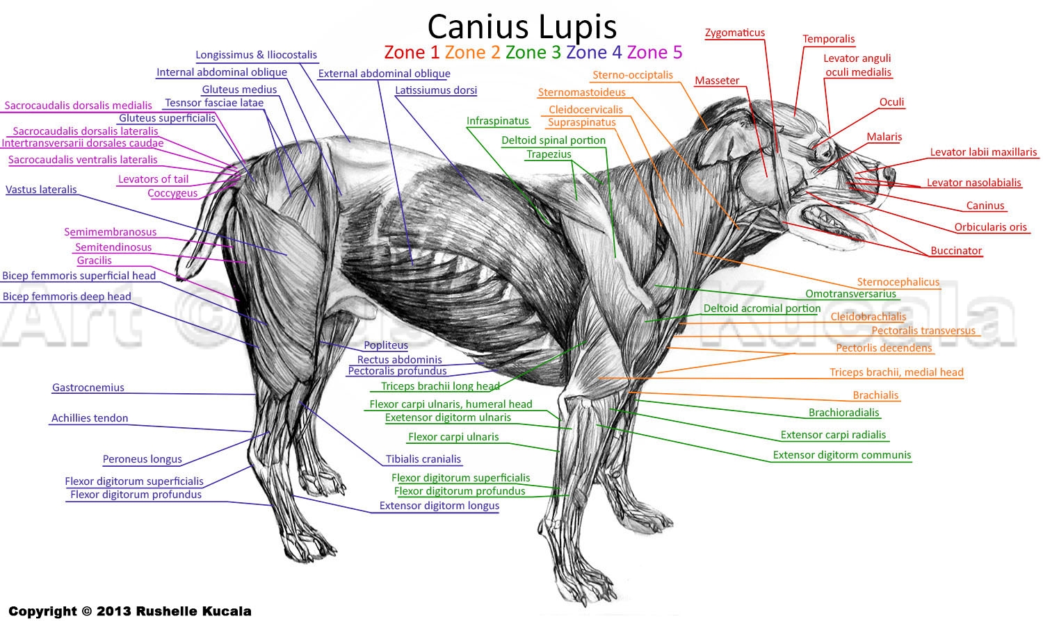
The major leg muscles in a dog play a crucial role in ensuring locomotion, stability, and flexibility. To understand their structure and function, it is essential to address the bones, joints, and ligaments that make up the dog's leg. In terms of bones, the dog's hindlimb mainly consists of the femur, the longest bone in a dog's body, which.
What Part Of A Dogs Leg Is The Knee
Dog anatomy comprises the anatomical studies of the visible parts of the body of a domestic dog.Details of structures vary tremendously from breed to breed, more than in any other animal species, wild or domesticated, as dogs are highly variable in height and weight. The smallest known adult dog was a Yorkshire Terrier that stood only 6.3 cm (2.5 in) at the shoulder, 9.5 cm (3.7 in) in length.
Dog Leg Anatomy in Human Speak Ortho Dog
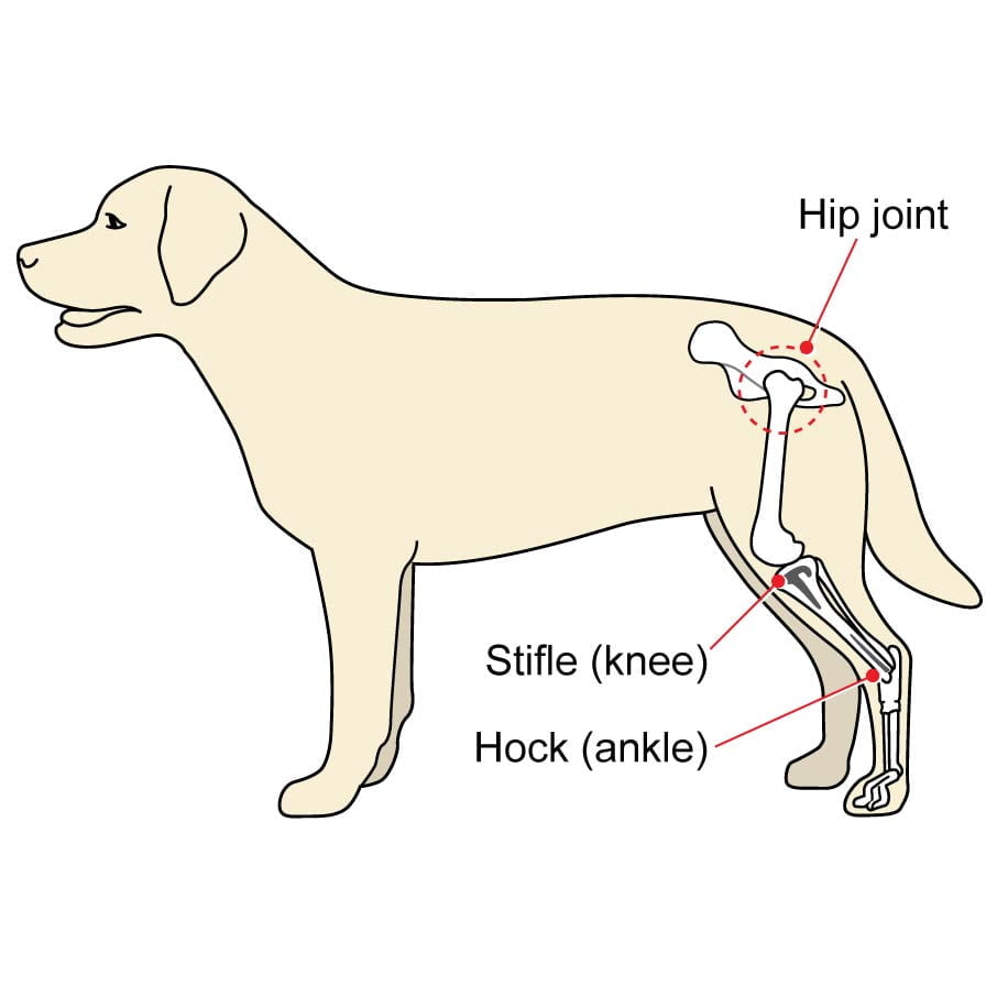
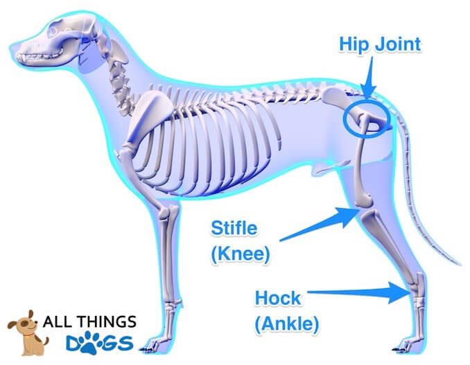
Dog Hind Leg Anatomy. The hind leg can be confusing to some owners, but it has some of the same features as a human. The bone between the hip and knee is the femur. Below the knee is the tibia and fibula. Then we get to the hock; you've probably heard this mentioned more in horses. The hock is like the human ankle.

Best Guardian Dog Rottweiler's Anatomy Dog anatomy, Rottweiler, Dog leg
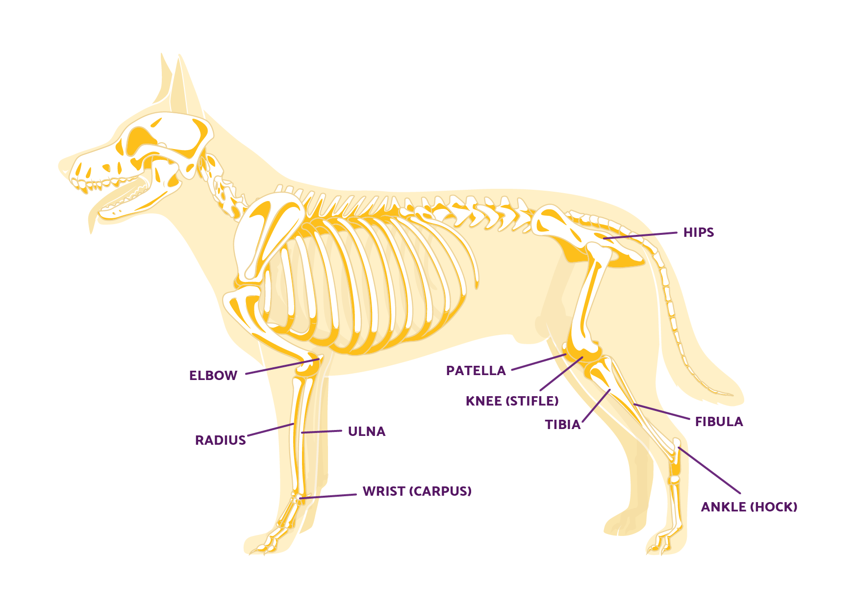
Dog rear leg anatomy. A dog's rear legs are where the largest bones and muscles are found. Just like humans, dogs actually have knees, and these are located where the tibia and fibula meet with the femur (where the lower leg meets with the thigh in dog back leg anatomy). The knee joint is known as the stifle, while at the rear back of the leg.
Dog Muscle Anatomy by TheDragonofDoom on DeviantArt
Dog skeleton. As with any vertebrate animal, the skeleton of a dog has the function of supporting the body for movement and protecting its internal organs. We can divide the canine skeleton into three main sections: Axial skeleton: skull, spine, ribs and sternum bones. Appendicular skeleton: bones of the extremities.

22 best Animal Anatomy Canines images on Pinterest Animal anatomy
The dog leg anatomy is a complex structure composed of various key components that work together to support the dog's movement and overall functionality. Understanding these key structures can give valuable insights into the dog's physical abilities and any potential issues they may face.

Dog Leg Anatomy Dog anatomy, Dog leg, Dogs
The extensor digit I longus is a small muscle that directly lies on the tibia. This extensor muscle of the dog leg anatomy arises from the cranial border of the fibula bone between the proximal and middle third. The extensor digit I longus muscle extends obliquely deep to the long digital extensor, fibularis brevis, and finally, on the tibia bone.

Dog Leg Bones Diagram Bone broth is a great way to add valuable
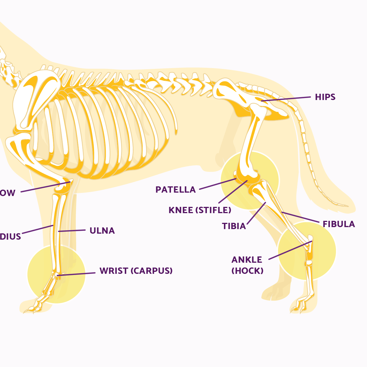
Dog Knee and Knee Cap. Dog leg anatomy is complex, especially dog knees, which are found on the hind legs. The technical term for a dog knee is the stifle joint. The stifle joint connects the femur, which is the dog thigh bone, to the tibia and fibula, the lower leg bones, and the patella,the canine equivalent to the knee cap. Many dogs.
A Visual Guide to Dog Anatomy (Muscle, Organ & Skeletal Drawings) All
Lameness is usually the first sign of a dog sprained leg — this indicates your dog is in pain. Swelling may follow. If the leg becomes warm or hot, however, it's unlikely that the limping is due to a simple sprain. If the sprain goes unaddressed, lameness may continue and, over time, leg muscles may weaken. When atrophy occurs in one limb.

biomedicalephemera Medial and caudal views of the musculoskeletal
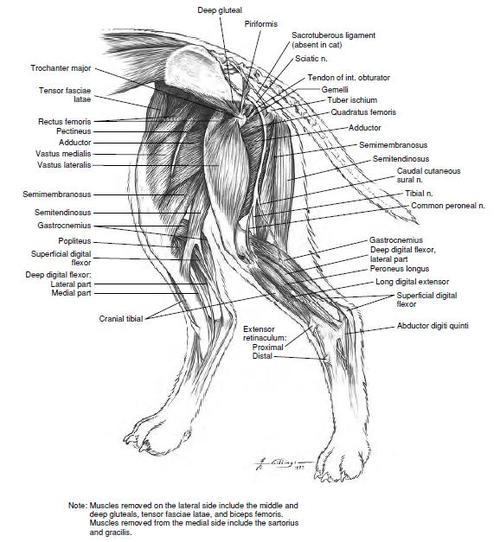
Or you may read the below-mentioned article to get a basic idea of the dog leg muscles - Dog leg anatomy with a labeled diagram (with bone, muscles, and vessels), Tibialis cranialis and dog's hock. The most superficial and flattened muscle lies cranial to the tibia bone. This tibialis cranialis muscle arises from the cranial part of the.
Dog Leg Anatomy in Human Speak Ortho Dog
Most first-year veterinary students have a misconception of the term "leg." Anatomically, the term leg means the part of the hind limb that extends from the stiffle joint to the hock joint (knee to ankle or tibia and fibula bones region). This short post will try to cover thedog leg anatomyin detail.

Dog anatomy front legs Canines Pinterest Dog anatomy, Anatomy
Dog anatomy lateral skeleton. The basics of the bones are: The femur: part of the dog's leg situated above the knee on the hind leg; The stifle or knee: the joint that sits on the front of the hind leg in line with the abdomen; Tibia and fibula: the part of the hind leg beneath the knee to the hock;

canine muscular anatomy Dog Muscles Diagram
Thighs: The upper thigh is above the knee of the hind leg. The lower thigh is beneath the knee. Stifle: The stifle, or knee, sits on the front of the hind leg. It falls in alignment with the abdomen. Hock: The hock is also known as the harsus. This is the joint on the dog's hind legs that makes an awkward sharp angle.

dogfrontlegboneanatomy Dog anatomy, Feline anatomy, Lion anatomy
Dog leg bone anatomy You know, the dog leg bone consists of tibia and fibula bones. In this part of the article, I will describe the dog leg bone anatomy in detail. The tibia is a long, thick bone that lies in the medial part of the leg. The proximal half of the tibia is triangular and more massive than the distal part.
